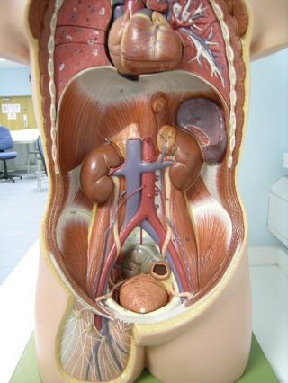
On Monday we spent the morning looking at the kidneys and suprarenal (or adrenal) glands. You saw their anatomy and histology in other stations, but with me we had a quick look at the anterior visceral relationships of each kidney, and then talked about the overall scheme and structure of the nervous system.
The kidneys lie between the peritoneum of the abdomen, and the musculature of the posterior abdominal wall. They are surrounded by fat and fascia, but the point I want to make here is that they are retroperitoneal. Some of the gastrointestinal tract and other abdominal organs are also retroperitoneal.
The right kidney is a little lower than the left kidney. The liver takes up a big space on the right hand side of the abdomen. The left kidney has the left suprarenal gland sat upon its superior pole, and the spleen lies a little laterally to this. Part of the stomach is also anterior to the superior pole of the left kidney. Anterior to the upper middle part of the left kidney lies the tail of the pancreas. This is also retroperitoneal. The left colic flexure (also known as the splenic flexure, because the spleen is here) of the large bowel lies anterior to the left kidney, along with loops of small bowel (jejunum).
The right kidney also has a suprarenal gland upon its superior pole, and the liver is here too. The descending duodenum runs along the medial part of the anterior surface of the right kidney, and is also retroperitoneal. Inferiorly the right colic flexure (or hepatic flexure) and loops of small bowel lie anterior to the kidney too.
We built this up using the plastic torso models, and you might want to do this again yourself. Try to imagine where the peritoneum covers the posterior abdominal wall, and where it reflects up to surround abdominal structures.
The nervous system.
At this stage (week 7) it is useful to have an overview of the anatomy of the nervous system. You've been hearing and using lots of terms for various parts in recent weeks, so let's tie it all together to see if you have developed a good feel for the system.
We can divide the nervous system into two parts: somatic and visceral. The word "somatic" is derived from the Greek word, "soma", meaning "the body". It refers to the frame of the body, rather than to the organs. So the somatic part of the nervous system refers to the parts of the body under voluntary control, largely skeletal muscles. When I described this I tried to get you thinking in motor terms only, for simplicity, but the somatic part of the nervous system includes neurones involved in the sensory input that keeps the body in touch with its surroundings, i.e. external sensory input from the skin, sight and sound.
So the visceral part of the nervous system must drive and receive sensory information from everything else. Smooth muscle and cardiac muscle are innervated by nerves of the visceral nervous system, as are the organs (the viscera), and sensory (afferent) fibres conveying noxious stimuli (pain) and other sensory information back to the central nervous system. You might want to call these visceral sensory neurones, "general visceral afferent fibres". This is all under unconscious, or involuntary control, so this part of the nervous system is more often referred to as the autonomic nervous system. The autonomic nervous system is further divided into the sympathetic and parasympathetic nervous systems.
Jumping back a bit, we can also divide the nervous system into the central nervous system (the brain and spinal cord) and the peripheral nervous system (nerves coming out of or going back into the central nervous system). The somatic and visceral parts of the nervous system are generally regarded as sub-divisions of the peripheral nervous system, but when you look at the brain and spinal cord anatomically next year you will see that this view is not always as clear cut as you might like it to be. We'll save that for later though. It's good fun.
Back to the autonomic nervous system: the sympathetic part of the nervous system is often described as triggering the "fight or flight response". If a terrifying cardiothoracic surgeon pounces on you with questions about a coronary angiogram that you struggle to understand, your body may respond by reducing blood flow to organs that aren't useful right now, diverting blood flow to skeletal muscles and releasing glucose into the blood from stored glycogen, preparing you to strike the surgeon (probably not the best option) or flee. This is the sympathetic nervous system acting. Interestingly, as you looked at the suprarenal glands this week, the medulla of this gland contains chromaffin cells that are hard wired directly to sympathetic neurones. These neurones instruct the chromaffin cells to release adrenaline into the bloodstream, triggering body-wide fight or flight responses. Sympathetic neurones are found all over the body, as they drive vasoconstriction, including blood vessels of the skin.
The parasympathetic part of the nervous system is often mentioned as being involved with "rest and digest" functions. Sometimes the parasympathetic and sympathetic parts of the nervous system work in opposition, but often they have somewhat separate functions. Bear this in mind when you are trying to understand their effects on structures and systems in different parts of the body. Parasympathetic neurones will increase mucosal gland and salivary gland activity, increase intestinal activity, for example, but it will also slow the heart rate, whereas the sympathetic nervous system increases the heart rate.
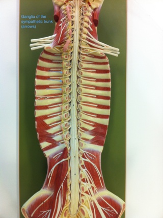
We talked a bit about the anatomy of the autonomic nervous system. We looked at how the sympathetic nerves leave the spinal cord and pass to the sympathetic trunk, two chains of ganglia (ganglion = collection of nerve cell bodies) running on either side of the vertebral column from the pelvis to the neck. The trick here is that sympathetic nerves only leave the spinal cord from levels T1 to L2, but once in the sympathetic trunk they can ascend or descend a little way before leaving to reach other parts of the body. Before they leave the sympathetic trunk most neurones will synapse with another neurone in a ganglion. So we call the fibres going into the sympathetic trunk "preganglionic sympathetic neurones", and those leaving (a second neurone that has synapsed with the first and received the action potential) "postganglionic sympathetic neurones".
If sympathetic nerves leave the spinal cord in the middle (thoracic and upper lumbar segments), then parasympathetic nerves leave the central nervous system at either end; that is, some cranial nerves coming out of the brain contain parasympathetic neurones, and spinal nerves of the sacral plexus contain parasympathetic neurones that enter the pelvis. You've seen how parasympathetic innervation gets into the thorax and most of the abdomen: the vagus nerve (cranial nerve X) descends in the neck and runs through both the thorax and the abdomen.
If a (nerve) ganglion is collection of nerve cell bodies (synapsing with incoming axons of other neurones) what is a nerve plexus? A plexus is a collection of nerve fibres running together, crossing over, changing direction, and maybe forming new, larger nerves. There are no synapses here. There's no informational exchange between nerve fibres in a plexus. Think of it more as a collection of cables that are being organised with cable tidies (thanks for that idea, year 1).
On the abdominal aorta we find both ganglia and plexuses. Near the branch of the coeliac trunk from the aorta we find a couple of masses called the coeliac ganglia, and a meshwork of autonomic nerve fibres running away with the major arteries to pass to the viscera of the abdomen. Going back to the kidney, we might say that the kidney receives autonomic nerve fibres from the aorticrenal plexus (see the parts of the name in there). Sympathetic neurones are vasomotor to the afferent and efferent arterioles within the kidney, but it's unclear what role parasympathetic innervation may play here. Regulation of kidney function is predominantly influenced by hormones.
When we look at the abdomen again in future sessions, and when we cover the embryology of the endocrine system, you will start to add to these ideas of autonomic innervation. Hopefully with time you'll build up a picture of the wiring of the gastrointestinal system and related organs.
Tweet


Comments